Overview
The Widom Lab studies RNA, which performs a plethora of different functions in the cell by folding into specific structures and, often, interconverting between different structures. In addition to its well-known roles as an intermediate messenger between DNA and proteins, RNA is also capable of regulating gene expression, catalyzing chemical reactions, and much more. Our lab is particularly interested in riboswitches, which regulate gene expression in bacteria, and splicing, the process by which sections of RNA that do not code for proteins are excised.

We use a variety of biophysical techniques, centered around ultrafast and single-molecule spectroscopy, to probe the structures and dynamics of systems ranging from isolated RNAs to large RNA-protein complexes.
Current Work
We are utilizing steady state fluorescence and circular dichroism spectroscopy to study local conformational changes that occur upon folding of riboswitches. For these experiments, we utilize the fluorescent nucleic acid base analogues (FBAs) 2-aminopurine (2-AP) and pyrrolocytosine (pC). When 2-AP is incorporated into RNA, its fluorescence gets quenched, while the opposite occurs for pC. In both cases, the CD spectrum of the RNA gets modified as a result of electronic interactions between the FBA and its neighboring bases (right, panel A). These changes can be used to analyze the folding mechanism of the RNA and the impacts of ligand binding. We are also utilizing 2-AP as a donor fluorophore for Förster resonance energy transfer (FRET) experiments in order to probe nanometer length-scale structural rearrangements in the RNA (B).
FRET can be measured on single molecules if dyes that are brighter than 2-AP and pC are used. We are using single-molecule FRET and other single-molecule methods to probe conformational rearrangements in a variety of RNA, including riboswitches (C), transfer RNA and long noncoding RNA. We also utilize biolayer interferometry to analyze the binding of ligands and proteins to RNA (D). For example, we showed that a riboswitch labeled with two fluorophores did not bind ligand (cyan) while a riboswitch labeled with one fluorophore did (red).


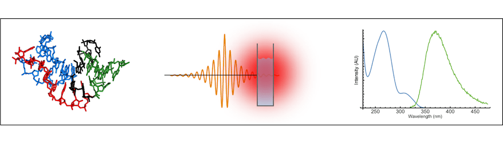
In addition, we have built an instrument that measures excited state lifetimes on an ultrafast time-scale. We are using this instrument to investigate fluorescence quenching and energy transfer in 2-AP and other probes in a far more nuanced way than simply looking at their fluorescence intensities.
Publications
Coulson, T. L.; Widom, J. R.* “Impacts of sequence and structure on pyrrolocytosine fluorescence in RNA”. Nucleic Acids Res., 2025, 53, gkaf262. Link
Conoan Nieves, N. E.; Widom, J. R.* “Ligand binding characteristics of an NAD+ riboswitch revealed by FRET and biolayer interferometry”. Chem. Comm., 2025, 61, 346-349. Link
Hoeher, J. E.; Sande, N. E.; Widom, J. R.* “Probing and Perturbing Riboswitch Folding using a fluorescent base analogue”. Photochem. Photobiol., 2024, 100, 419-433. Link
Wonderlick, D. R.; Widom, J. R.; Harms, M. J.* “Disentangling contact and ensemble epistasis in a riboswitch”. Biophys J. 2023, 122, 1600-1612. Link
Widom, J. R.*; Hoeher, J. E. “Base-stacking heterogeneity in RNA resolved by fluorescence-detected circular dichroism spectroscopy”. J. Phys. Chem. Lett., 2022, 13, 8010-8018. Link